Hugh Kim & Dongjoon Im
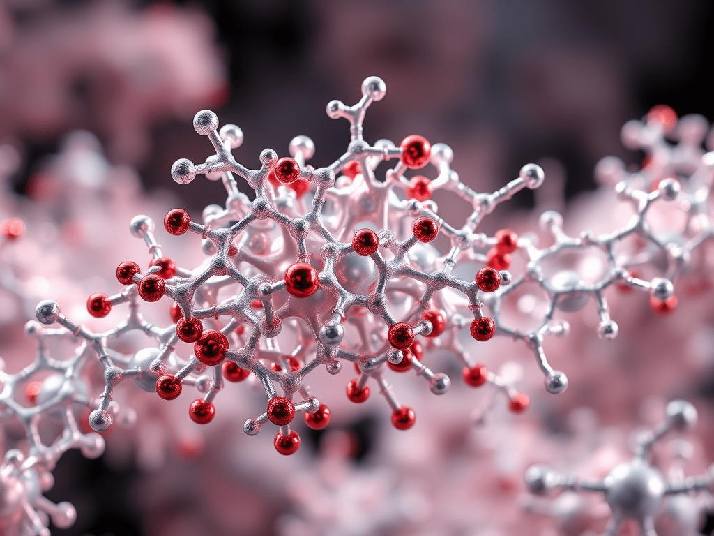
The most prominent amyloid protein is β-amyloid peptide 1-42 (Aβ42). Alzheimer’s Disease (AD), the most common form of dementia, is characterized by the accumulation of neurofibrillary tangles and amyloid plaques, which are aggregates of microtubule-associated protein tau and β-amyloid peptides (Aβ), respectively. Numerous studies have identified the cleavage of amyloid precursor protein (APP) into the neurotoxic Aβ42 as a major cause of AD. APP, located in the cell membrane, is highly expressed in the brain and is processed into Aβ peptides by the enzymes β-secretase and γ-secretase. Aβ peptides can vary in size, ranging from 37 to 43 amino acids, with Aβ42 specifically containing 42 amino acids.
In 1984, the amino acid sequence of Aβ peptides was first identified in amyloid plaques deposited outside cortical cells. Aβ40 and Aβ42 are the major components of these plaques. Although the function of the Aβ peptide remains unclear, its pathological aggregates are well-known to be associated with Alzheimer’s. Research indicates that the cellular toxicity of early-stage Aβ oligomers exceeds that of mature β-sheet-rich amyloid fibrils. Among Aβ peptides, Aβ40 is the most prevalent, but Aβ42 exhibits the greatest toxicity due to its rapid amyloid fibrillation and high nucleation rates during early aggregation.
Amyloid plaques contain metal ions such as copper, iron, and zinc, which coordinate with Aβ42 to promote fibrillation. These metals, upon forming coordination bonds with Aβ42, expose hydrophobic regions, accelerating hydrophobic interactions between Aβ42 peptides. Some studies suggest that high concentrations of zinc or copper induce amorphous aggregation, potentially inhibiting the amyloid fibrillation of Aβ peptides.

Although Aβ40 undergoes amyloid fibrillation, its aggregation rate is much slower than that of Aβ42. Aβ40 differs from Aβ42 by lacking two C-terminal hydrophobic amino acids (isoleucine and alanine), which are critical in stabilizing hydrophobic interactions within the fibril. While the thermodynamic role of these residues in fibril stabilization is understood, their kinetic role in accelerating fibrillation remains unclear. Studies using ion mobility mass spectrometry reveal that Aβ40 and Aβ42 form distinct tetrameric oligomers, leading to faster nucleation and toxicity in Aβ42. Interestingly, these peptides rarely co-aggregate into fibrils, likely due to differences in their oligomerization dynamics.
Another hallmark of AD is the formation of intracellular neurofibrillary tangles in the medial temporal lobe, caused by amyloid fibrillation of tau protein. Tau stabilizes microtubules and supports neuronal structure and differentiation. Tau, a basic protein with a net positive charge at neutral pH (pH 7), interacts electrostatically with microtubules. However, this charge also creates a barrier to amyloid aggregation due to repulsive forces between positively charged residues. Hyperphosphorylation of tau, a critical mechanism in its detachment from microtubules and fibrillation, remains poorly understood. The negatively charged phosphate groups overcome electrostatic repulsion, facilitating tau aggregation.

Negatively charged biological molecules such as ATP and RNA promote tau aggregation by reducing electrostatic repulsion. Recent studies suggest that ATP induces tau aggregation without forming part of the fibrils, implying stronger stabilizing hydrogen bonds in β-sheet structures compared to electrostatic repulsion from lysine residues.
Both Aβ and tau aggregation are central to Alzheimer’s pathology. However, their distinct locations—Aβ outside cells and tau inside—result in different behaviors and interaction ranges. Although their interaction has been proposed, its exact nature remains elusive. Recent research suggests that tau may delay the oligomerization of Aβ, but the precise mechanisms are unknown. Understanding their aggregation pathways is crucial for developing treatments for neurodegenerative diseases.
Please visit the Hugh Kim Research Group homepage.
Refernces
- Goedert et al. Neuron 1989, 3(4), 519-526
- Hardy, J. A. and Higgins, G. A. Science 1992, 256(5054), 184-186
- Chen et al. Acta Pharmacol Sin 2017, 38(9), 1205-1235
- Glenner, G. G. and Wong, C.W. Biochem Biophys Res Commun 1984, 120(3), 885-890
- Uversky et al. J Biol Chem 2001, 276(47), 44284-44296
- Choi et al. J Am Chem Soc 2017, 139(43), 15437-15445
- Leal et al. Coord Chem Rev 2012, 256(19-20), 2253-2270
- Gu, L. and Guo, Z. J Neurochem 2013, 126(3), 305-311
- Gremer et al. Science 2017, 358(6359), 116-119
- Bernstein et al. Nat Chem 2009, 1(4), 326-331
- Heo et al. Int J Mass Spectrom 2018, 428, 15-21
- Carlomagno et al. Cell Rep 2021, 34(11), 108843
- Wallin et al. J Am Chem Soc 2018, 140(26), 8138-8146

Leave a comment